Nuclear Medicine and Molecular Imaging
Nuclear Medicine is the specialty of Radiology that uses radioactive tracers (radiopharmaceuticals) to diagnose and treat a variety of diseases. Unlike most other imaging, Nuclear Medicine identifies how the body functions. A large selection of radiopharmaceuticals allows physicians to identify abnormalities in many different organ systems. In larger amounts, these radiopharmaceuticals can even be used to treat several different cancers.
Molecular Imaging is the next step in the evolution for Nuclear Medicine and builds on the tracer principles for investigating disease processes. These tracers are advanced radiopharmaceuticals that explore diseases at the cellular and molecular level. Antibodies, peptides and other molecules target the cellular functions and metabolic pathways of the cells and tissues. A blend of recent scientific advances in molecular biology, chemistry and physics allows the physicians to investigate disease processes in ways that complement the rest of Radiology and provide unique information. Combined with the latest technology of PET/CT and SPECT/CT, abnormalities can be identified earlier and localized like never before.
MISSION
The staff of the Division of Nuclear Medicine and Molecular Imaging is dedicated to providing the referring physician with clear and critical information on a timely basis in a safe, friendly and pleasant environment. We employ dose-reduction strategies to minimize radiation exposure and maximize image quality. As part of our educational mission, we train the next generation of medical students, residents, and fellows in the techniques of functional imaging while exploring research opportunities.
CLINICAL SERVICE
The division is equipped with five dual-headed gamma cameras. Attenuation correction is employed for SPECT studies to improve quality and decrease artifacts. SPECT/CT allows the fusion of functional and anatomic data. A PET and PET/CT scanner provides essential information in the management of cancer, heart disease and neurologic disorders like seizures. The HERMES software suite provides us with the ability to merge quickly new functional images with prior CT and MR imagery, providing three-dimensional imaging, quantification and localization. A portable gamma camera allows for imaging of critically ill patients while they are in the intensive care unit. An in-vitro lab is also available to do several non-imaging studies. Services are provided at Loyola University Medical Center and Gottlieb Memorial Hospital.
Nuclear Medicine and Molecular Imaging collaborates with physicians across many specialties throughout the institution. For example, the team works closely with endocrinologists in the treatment planning of hyperthyroidism and thyroid cancer; provides data on the effectiveness of chemotherapy to the oncologists and identifies the presence and location of cardiac disease for the cardiologists. Collaboration with the interventional radiologists allows for the selective treatment of liver tumors. For neurologists and neurosurgeons, seizure foci can be identified as part of planning for surgical therapy. Radiation oncologists benefit from tumor localization as a part of treatment planning and follow up.
EDUCATION
The physicians, scientists and technologists are available to speak to other professionals or to the public on a variety of topics pertaining to Nuclear Medicine and Molecular Imaging, including: radiation and radiation safety, equipment performance and quality control. Our residency program admits one person each year to complete a three-year training program that emphasizes the multidisciplinary diagnostic and therapeutic aspects of Nuclear Medicine and Molecular Imaging. The training program includes rotations through CT, MRI, neuroradiology, medical oncology and radiation oncology. A one-week training session at Oak Ridge National Laboratories at the REAC/TS facility provides training on proper response and management of accidents involving radioactive materials.
RESEARCH
Some members of the division have developed additional expertise or board certification based on their interests. These include the Certification Board of Nuclear Cardiology and PET/CT training offered by the American College of Radiology. There is active research that has been approved by the Institutional Research Board for the Protection of Human Subjects. Additional projects are planned for the future. Areas of research interest include radiation biology, gamma-specific breast imaging, the management of radiation accidents and quality assurance mechanisms for imaging equipment.

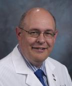
Robert Wagner M.D., FACR, FACNP
Professor
Medical Director, Nuclear Medicine
Radiology
Specialties
- Computers in Medicine
- Molecular Imaging
- Nuclear Cardiology
- PET and F-18 Imaging
- Radiation Accidents and Biological Effects
- Nuclear Medicine
